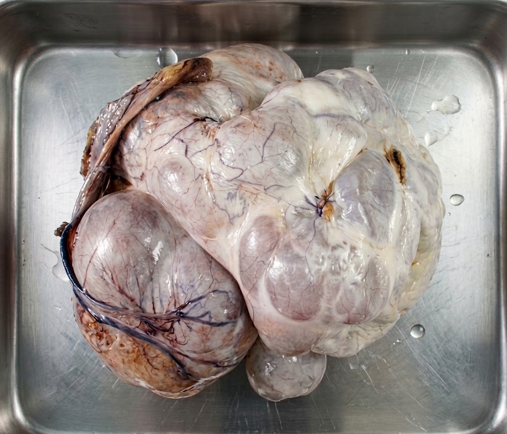
A Case Report. 40 years old pleasant patient walks into the clinic with a referral from general practitioner for pelvic mass. Her reason for the visit to the GP clinic was mild abdominal discomfort. She did not experience any abdominal pain nor did she realize the gross abdominal distention. Her menstrual cycles were regular with occasional slight dysmenorrhea. Despite having mild indigestion issues, she did not complain of loss of weight or appetite. Her weight was a mere 51kg at the initial consult. She gave no history of gynaecological malignancy in the family. On examination, there was no pallor, her vitals were normal and lungs were clear. Thyroid and breast were unremarkable. Her abdomen however was grossly distended with a hard mass occupying it in entirety up to the xiphisternum. Routine blood works and tumour markers were normal. CT abd- pelvis revealed a large multicystic mass with soft tissue and solid component arising from pelvis measuring 18×25.6x29cm. No enlarged pelvic or para-aortic lymph nodes seen. No ascites or omental mass noted as well. After counselling the patient, she opted for a unilateral salpingo-oohorectomy with the knowledge of further surgery pending the hIstopatahological result. A midline laparotomy was performed. Intraoperatively, there was a large left ovarian tumour which was well encapsulated and as planned, a left SO was done. Rest of abdomen was unremarkable. She recovered well post operatively and was discharged on day 3 of surgery. The mass weighed 3845gm and the Histopathological exam revealed : i) Mature cystic teratoma. No immature component or malignancy. ii) Mucinous borderline tumour. No stromal invasion. iii) Peritoneal cytology- no malignant cells. Discussion Large ovarian tumours are rare occurances. Benign mucinous cyst adenomas account for about 15% of all ovarian neoplasms and are among the largest known tumours. About 80% of mucinous tumours are benign, 10% are borderline and 10% are malignant. They are usually common between third and fifth decade of life. The treatment of choice is surgery. It can be accomplished by en bloc removal of tumour with or without controlled drainage of tumour fluid. Removal of tumour en bloc is thought to decrease the risk of spilling potentially malignant cells. However controlled drainage prevents supine hypotension syndrome . Borderline ovarian tumors (BOTs) comprise 10–20% of ovarian malignancies with peculiarity of absence of clinical manifestation. Although they are similar to malignant epithelial ovarian tumors in some of the histologic characteristics, these types of tumors do not have destructive stromal invasion. Their prognosis is much better than that of carcinoma. These mucinous tumors usually appear as large cystic multilocular masses containing sticky gelatinous fluid. Most of them are unilateral, well-differentiated and, when diagnosed in Stage I, present a recurrence rate of 1%. They appear to cause compressive symptoms to the adjacent structures in the abdomen due to their sheer size and rarely any hormonal symptoms. Operative risk is extremely high, with fatal complications described in literature , i.e- pulmonary and cardiac failure, pulmonary embolism and sepsis. These are related to sudden changes of intra-abdominal pressure, during surgery and in postoperative phase. In conclusion, giant ovarian tumors are rare neoplasms which needs careful treatment, preventing high-risk operative complications. Symptoms at diagnosis is related with prognosis, while asymptomatic cases are not often related with better results. Dr. Prema Latha Supramaniam Consultant Obstetrician and Gynaecologist KPJ Rawang Specialist Hospital.